
Scintillators represent remarkable materials that illuminate when exposed to high-energy particles or X-rays. Within medical and dental imaging systems, these sophisticated components transform incoming X-ray radiation into visible light that can be captured using film or digital photosensors. Beyond healthcare applications, scintillators play crucial roles in night-vision technologies and various research environments, including particle detectors and advanced electron microscopes.
In a groundbreaking development, researchers at MIT have demonstrated how to enhance scintillator efficiency by at least tenfold, with potential improvements reaching up to a hundredfold. This revolutionary advancement comes through engineering the material's surface to create specific nanoscale configurations, including arrays of wave-like ridges. Unlike previous approaches that focused primarily on discovering new materials, this innovative methodology could theoretically be applied to any existing scintillator materials.
While integrating these advanced scintillators into current X-ray machines will require additional time and development, the research team anticipates significant improvements in medical diagnostic X-rays and CT scans. These enhancements could substantially reduce radiation exposure while simultaneously improving image quality. In industrial applications, such as X-ray inspection of manufactured components for quality assurance, the new scintillator technology could enable more precise inspections conducted at higher speeds.
The findings were detailed today in the prestigious journal Science, in a paper authored by MIT doctoral students Charles Roques-Carmes and Nicholas Rivera; MIT professors Marin Soljacic, Steven Johnson, and John Joannopoulos; along with ten additional collaborators.
Although scintillators have been utilized for approximately 70 years, most research in this field has concentrated on developing new materials that produce brighter or faster light emissions. This novel approach instead applies cutting-edge nanotechnology advances to existing materials. By creating precise patterns in scintillator materials at a length scale comparable to the wavelengths of emitted light, the team discovered they could dramatically alter the material's optical properties.
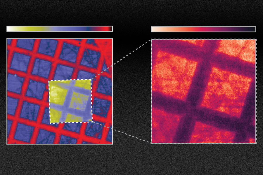
To create what they termed "nanophotonic scintillators," Roques-Carmes explains, "you can directly fabricate patterns within the scintillators, or you can attach another material containing nanoscale holes. The specific implementation depends on the exact structure and material composition." For this research, the team took a conventional scintillator and created holes spaced approximately one optical wavelength apart, or about 500 nanometers (billionths of a meter).
"The foundation of our work lies in the comprehensive theory and framework we've developed," Rivera notes. This enables the researchers to calculate the scintillation levels that would be produced by any arbitrary configuration of nanophotonic structures. The scintillation process itself involves a complex series of steps, making it challenging to fully understand. The framework developed by the team integrates three distinct types of physics, Roques-Carmes explains. Using this system, they've achieved excellent alignment between their theoretical predictions and experimental results.
The experiments demonstrated a tenfold improvement in emission from the modified scintillator. "This advancement could translate into significant benefits for medical imaging applications, which are typically photon-starved—meaning the conversion of X-rays to optical light limits image quality," Roques-Carmes states. "In medical contexts, minimizing patient exposure to X-rays is crucial, particularly for routine screenings and especially for pediatric patients."
"We believe this research will inaugurate an entirely new field within nanophotonics," he adds. "Researchers can leverage the extensive existing work in nanophotonics to substantially improve materials that exhibit scintillation properties."
"The research presented in this paper holds enormous significance," says Rajiv Gupta, chief of neuroradiology at Massachusetts General Hospital and an associate professor at Harvard Medical School, who was not involved in this study. "Nearly all detectors used in the $100 billion medical X-ray industry are indirect detectors—the exact type addressed by these new findings. Everything I use in my clinical practice today operates on this principle. This paper improves the efficiency of this process by tenfold. If this claim proves even partially true—say the improvement is two times instead of ten—it would be transformative for the field!"
Soljacic notes that while their experiments demonstrated a tenfold improvement in emission within specific systems, through further refinement of the nanoscale patterning design, "we've also shown that certain scintillator systems can achieve up to a hundredfold improvement, and we believe we've identified pathways toward even greater enhancements."
Soljacic highlights that in other nanophotonics areas—a field dealing with light-matter interactions at the nanometer scale—the development of computational simulations has enabled rapid, substantial improvements, particularly in solar cell and LED development. The new models this team created for scintillating materials could facilitate similar technological leaps, he suggests.
Nanophotonics techniques "provide the ultimate capability to tailor and enhance light behavior," Soljacic explains. "However, until now, applying this capability to scintillation remained unattainable because modeling the scintillation process was extremely challenging. This work, for the first time, fully opens the field of scintillation to nanophotonics techniques." More broadly, the team believes that combining nanophotonics with scintillators could eventually enable higher resolution imaging, reduced X-ray dosage, and energy-resolved X-ray imaging capabilities.
This research is "very original and excellent," according to Eli Yablonovitch, a professor of Electrical Engineering and Computer Sciences at the University of California at Berkeley, who was not involved in this study. "New scintillator concepts are tremendously important in medical imaging and fundamental research."
Yablonovitch adds that while the concept still requires validation in practical devices, he notes that, "After years of research on photonic crystals in optical communication and other fields, it's long overdue that photonic crystals be applied to scintillators—materials of great practical importance that have been surprisingly overlooked until this work."
The research team included Ali Ghorashi, Steven Kooi, Yi Yang, Zin Lin, Justin Beroz, Aviram Massuda, Jamison Sloan, and Nicolas Romeo at MIT; Yang Yu at Raith America, Inc.; and Ido Kaminer at Technion in Israel. The work received partial support from the U.S. Army Research Office and the U.S. Army Research Laboratory through the Institute for Soldier Nanotechnologies, the Air Force Office of Scientific Research, and a Mathworks Engineering Fellowship.

